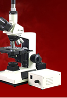
Examining the Structure of a Plant’s Root under a Microscope
The following microscope lesson and activity can be done by elementary and high school students or children. We will be examining the roots of a plant with the use of a high power compound light microscope. A high power kid’s toy microscope can also be used by children for this activity if they don’t have access to a higher grade student microscope.We all know that the roots function as the plant’s anchor to the ground. It supports the stem to maintain an upright position so that the leaves will get enough air and sunlight. The roots also absorb water from the soil together with the soil’s minerals that are essential to the food manufacturing process of the plant. In some cases, the roots also serve as the storage bin of the plant’s manufactured food. Examples of these are root crops like carrot and radishes.
To start with our microscope experiment, we first need to get a seedling of a grass with roots that contain a few hairs. We should mount this root on a microscope slide and add a drop of water. Under a high power compound light microscope, the root seems to be made up of numerous cells that are regularly arranged and in different shapes and sizes. The extreme tip of this structure is the root cap and if we focus our compound microscope on this thimble shaped tip, we will notice that the cells here are easily detachable. This is because these cells are worn off as the root pushes itself in the soil and is eventually replaced by new cells.
If we focus the science microscope at the upper part of these loose cells, we will notice that there is a region of small rectangular cells. These mass of cells are the new cells that are in the process of formation. As it matures, it grows longer until it becomes elongated cells above the small mass of cells. These elongated cells add length and thickness to the root.
Now we focus our examination to the fine hairs that extend from the root. These are the root hairs and under the high power compound microscope, they look like finger-like projections from the outer layer of the roots or the epidermis. You will also notice that these fine hairs have very thin walls. The thin walls allow the entry of water in the root and also of the salty minerals that the plant needs for its manufacture of food.
The next activity that we will do with the root is to examine what it looks like inside with the use of the compound light microscope. What we need to do is to cut a cross-section of the root (be sure to cut the hairy portion) very thinly that it is possible for light to pass through. An ideal root to be used is from that of a pea or been seedling.
Once we look at this sample under a high magnification childrens microscope, we will notice that it has numerous cells that are grouped together based on their appearance. Collectively, these cells are called tissues and they are divided into three: the epidermis, the cortex and the central cylinder or the stele.
You can easily recognize the epidermis under the compound light microscope because they are the cells at the outermost layer. The cells are tightly packed against each other so the only way that the water can enter the root is through them.
The majority of the root that we can see under the educational compound microscope is the cortex. The cortex serves as the water passage from the epidermis to the stele. It also sometimes functions as the food storage area of the plant like that of a turnip. As we focus our microscope towards the center of the root, we will notice that the stele cells thicken and form a ring around the innermost layer. This endodermis ring serves as a protective layer of the stele and also prevents the water from coming out.
Under the kid science microscope, the stele looks to have a cross-shaped section called the xylem. It is made up of thick long tubes that dissolve the nutrients absorbed by the roots while conducting it upward. In the stele you will also see clusters of smaller cells that are collectively called the phloem. These are also long tubes but with very thin walls and it functions as the root’s food conductors. The xylem and the phloem are then enclosed by a ring of cells called the pericycle. It is in the pericycle where the branch roots come from. If we look at the roots using a high power compound light microscope, we will notice that as we move upward, the cells differ in structure. This is because as the roots grow old, it becomes more and more like that of the stem until it completely assumes the function of the stem.
Studying about roots under high power microscopes helps students to learn more about nature that cannot be seen by their naked eye. It is also exciting to take something so seemingly basic and simple and realize that its structure and components are quite complex.


